MOHS MICROGRAPHIC SURGERY The Larkin Outpatient Multispecialty Center is proud to offer a new option for patients with skin cancer. Mohs Micrographic Surgery is a state-of-the-art technique used to remove recurrent and primary skin malignancies. This convenient outpatient procedure provides immediate results for cases characterized by -Removal of recurrent carcinoma of the skin -Tumors greater than two centimeters -Primary carcinoma (ears, nose, eyes, and lips) -Poorly-defined tumor borders -Incomplete excision of tumors -Tumors with aggressive growth patterns The procedure is performed by Dr. Jordan Fabrikant, who completed his residency at Larkin Community Hospital and now practices at the Larkin Outpatient Multispecialty Center. Mohs Surgery is named after its inventor, Dr. Frederick E. Mohs. It is an outpatient procedure in which the diseased area of the skin is removed through a series of microscopically controlled, shaved excisions. Mohs surgery has the highest cure rate of any other treatment for skin cancer. The outpatient procedure is cost-effective and convenient. The Mohs technique causes minimal damage to surrounding skin tissue, improving the likelihood of a positive cosmetic result. HOW IT WORKS The Mohs technique uses horizontal sections of the cancerous skin rather than conventional vertical incisions to ensure that the tumor is completely removed. A thin layer of the skin cancer is removed and examined under a microscope. If cancerous cells are detected, additional layers are removed. [caption id="attachment_8886" align="alignleft" width="626"] Scott Martini, Mohs Lab Technician prepares biological tissue for observation under a microscope, by cutting it in thin slices. Most biological tissue is too soft to cut; therefore, the tissue is frozen and sectioned in a cryostat as shown.[/caption] [caption id="attachment_8887" align="alignleft" width="627"]
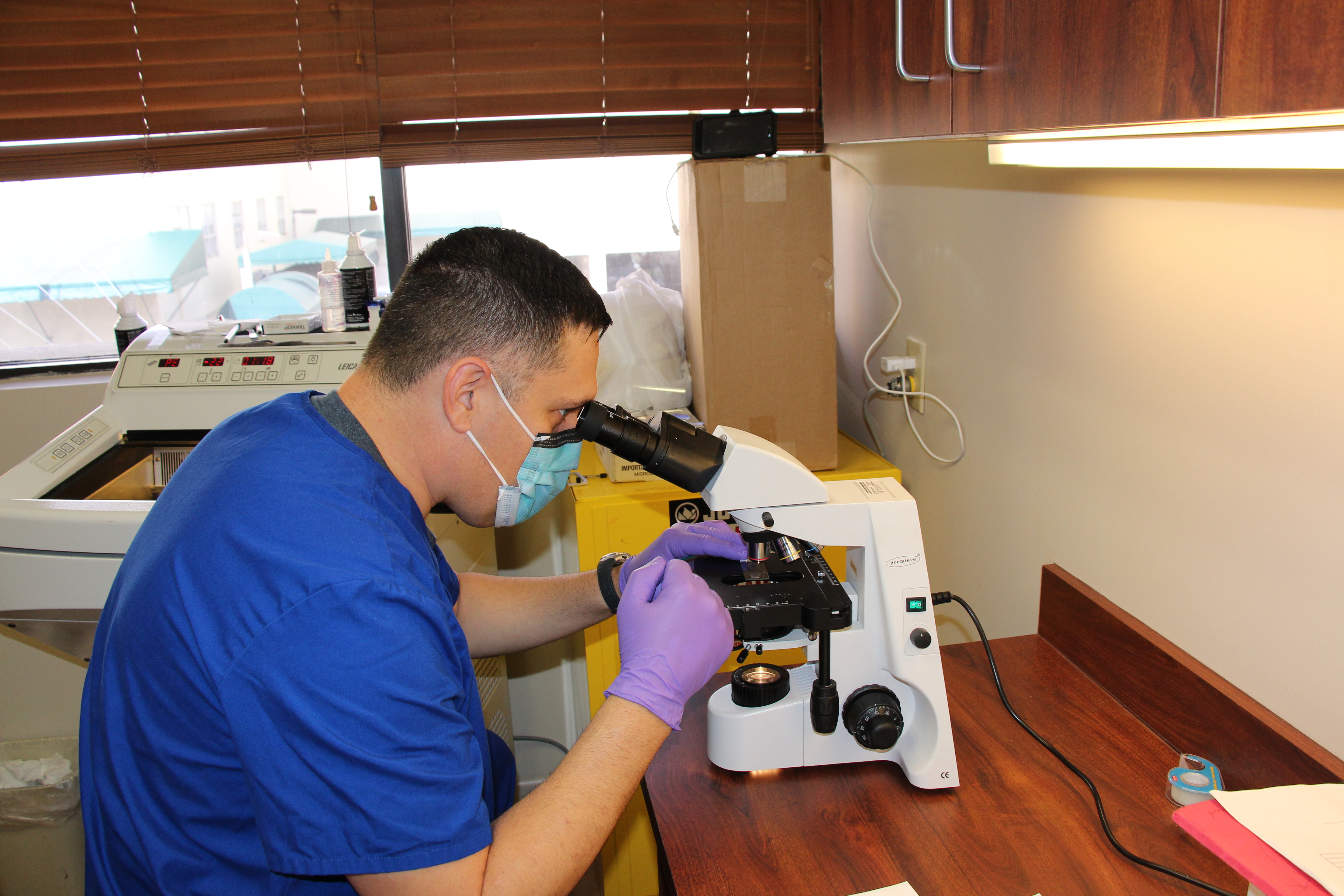 Scott Martini, Mohs Lab Technician observes the sectioned tissue under the microscope.[/caption]

LARKIN OFFERS STATE-OF-THE-ART TECHNIQUE TO REMOVE SKIN CANCER
Pingbacks
Pingbacks are closed.
Comments
Comments are closed.